Ear Wikipedia
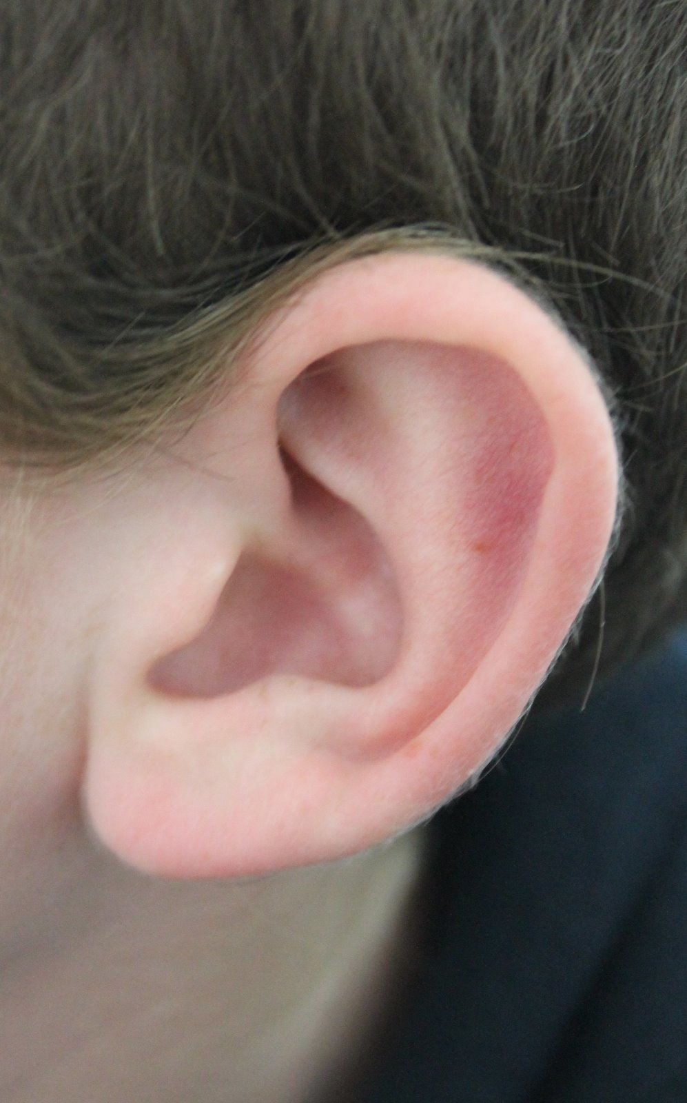
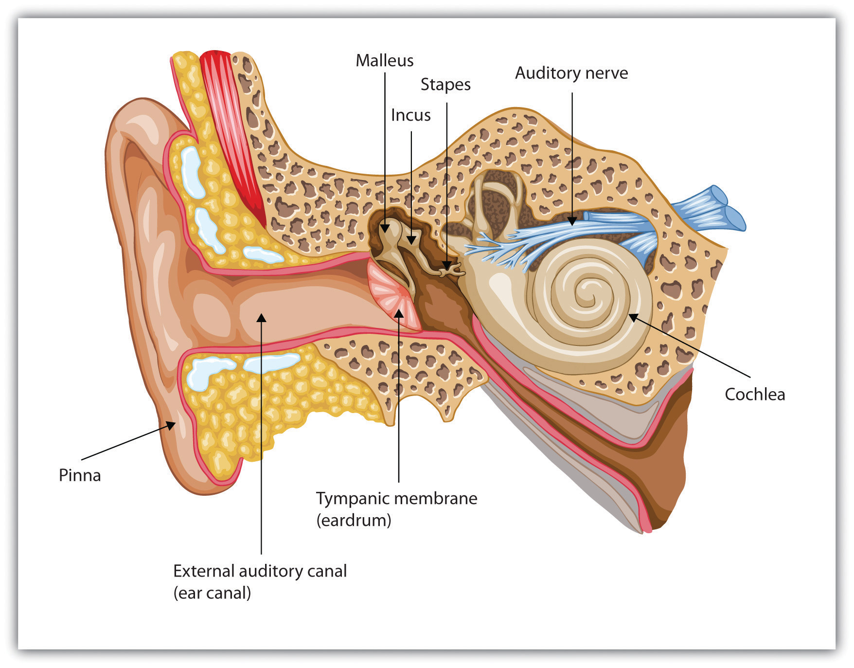
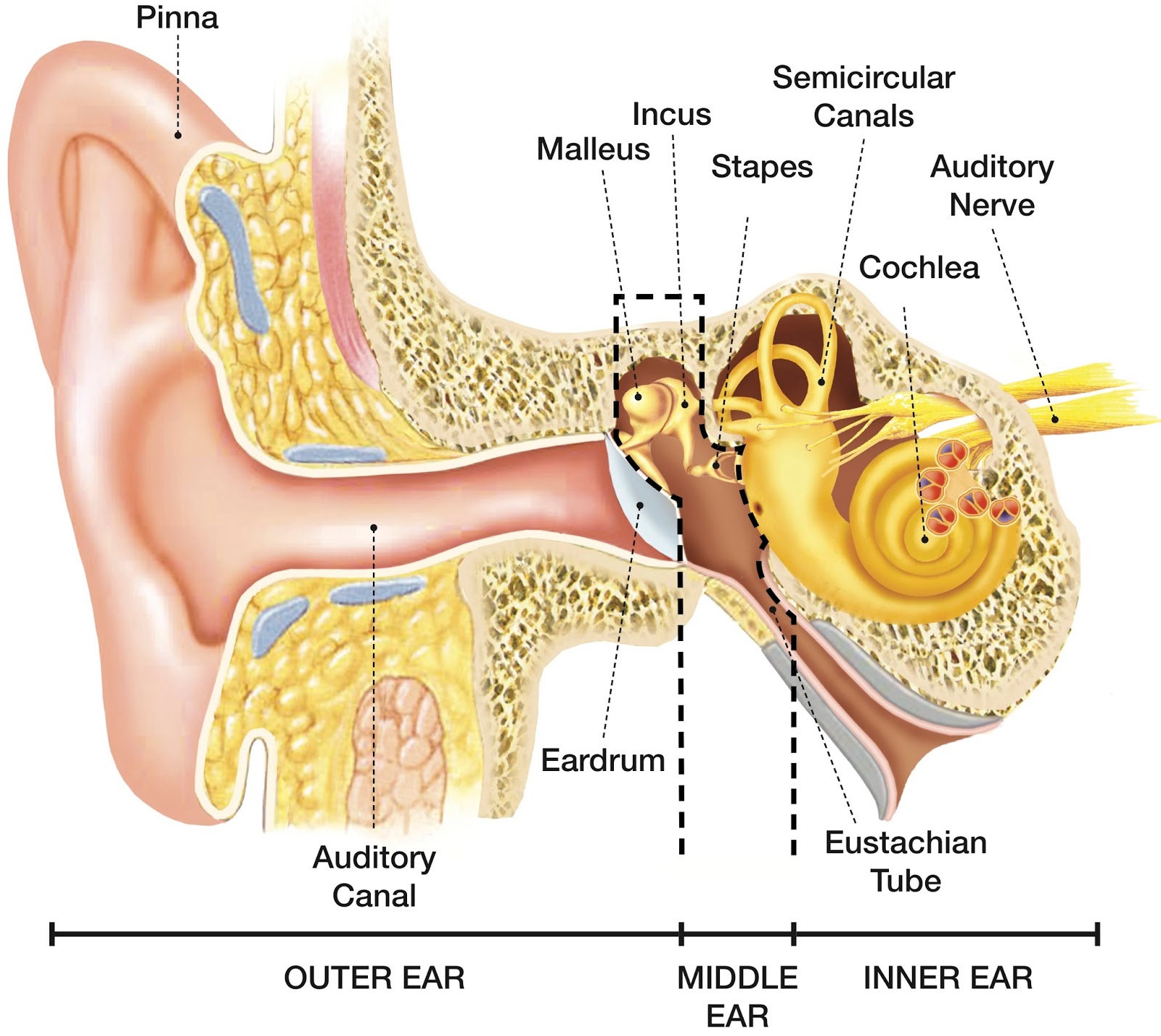
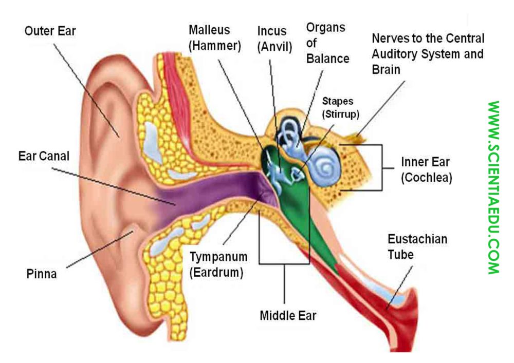
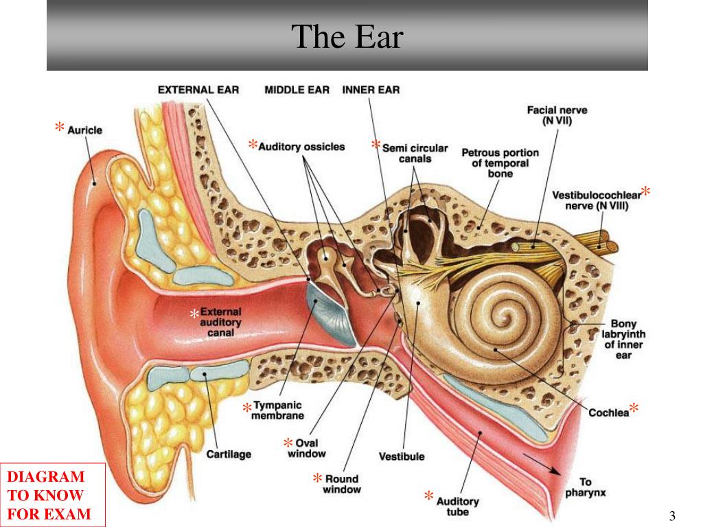
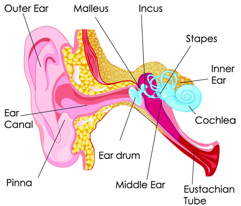
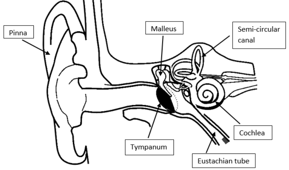
Ear anatomy. The external (outer) ear consists of the auricle, external auditory canal, and eardrum (Figure 1 and 2). The auricle or pinna is a flap of elastic cartilage shaped like the flared end of a trumpet and covered by skin. The rim of the auricle is the helix; the inferior portion is the lobule. Under the skin the outer one third of the.
5.3 Hearing Introduction to Psychology 1st Canadian Edition
Browse 7,900+ ear anatomy stock photos and images available, or search for anatomy model or muscle anatomy to find more great stock photos and pictures. anatomy model muscle anatomy anatomy infographic anatomy drawing body human ear anatomy inner ear anatomy cat ear anatomy dog ear anatomy outer ear anatomy ear anatomy illustration

31 Label The Ear Worksheet Labels 2021
File. : Anatomy of the Human Ear.svg. Size of this PNG preview of this SVG file: 512 × 389 pixels. Other resolutions: 316 × 240 pixels | 632 × 480 pixels | 1,011 × 768 pixels | 1,280 × 973 pixels | 2,560 × 1,945 pixels. Original file (SVG file, nominally 512 × 389 pixels, file size: 50 KB) Render this image in .

Anatomy of the Ear [4]. Download Scientific Diagram
Ear Definition. The ear is the organ found in animals which is designed to perceive sounds. Most animals have some sort of ear to perceive sounds, which are actually high-frequency vibrations caused by the movement of objects in the environment. The human ear picks up and interprets high-frequency vibrations of air, while the sound-sensing.

Anatomy Of Ear Labeled How We Perceive Sound Davidson Hearing Aid
Ear Anatomy 34 year old female with large posterior Tympanic Membrane perforation (Hole in the ear drum) The ear drum is often transparent and looks like a stretched piece of clear plastic. The drum is approximately the size of a dime.
Label the Ear
Ear Anatomy - Inner Ear. Next to the middle ear in the bone of the skull is a small compartment which contains the hearing and balance apparatus known as the inner ear. The inner ear has two main parts. The cochlea , which is the hearing portion, and the semicircular canals is the balance portion. The cochlea is shaped like a snail and is.
SPEECH LANGUAGE PATHOLOGY & AUDIOLOGY HEARING DISORDERS OF THE OUTER EAR
diagram of the anatomy of the human ear. Three ossicles: malleus, incus, and stapes (hammer, anvil, and stirrup). The ossicles directly couple sound energy from the ear drum to the oval window of the cochlea. Detailed illustration for educational, medical, biological, and scientific use. Ear anatomy diagram,vector.

Labeled Diagram Of the Ear Best Of Tape In Notebook 5 Mins 50 12 3
Here is a blank human ear diagram for you to label, so that you can memorize the different parts of this vitally necessary organ, for good.

DIAGRAM OF THE EAR Unmasa Dalha
A brief description of the human ear along with a well-labelled diagram is given below for reference. Well-Labelled Diagram of Ear The External ear or the outer ear consists of Pinna/auricle is the outermost section of the ear. The external auditory canal links the exterior ear to the inner or the middle ear.
Human Ear Home Tuition Guwahati Assam
Ear with Labels 3-D Model These models are useful for patient education, professional training and patient use. The interactive image below has a large file size and may take a long time to load on screen. For best results, download the file (18.6 MB) to your computer and open the original file on your desktop computer.

Anatomy and Analysis of the Ear Dr. Anil Shah Ears External ear
447 ear anatomy with labels stock photos, 3D objects, vectors, and illustrations are available royalty-free. See ear anatomy with labels stock video clips Filters All images Photos Vectors Illustrations 3D Objects Sort by Popular Stapedius muscle vector illustration. Labeled anatomical ear structure scheme.

Human Ear Diagram Without Labels Human Ear Diagram Without Labels Human
Stock Photos & High-Res Pictures. stock photos, high-res images, and pictures, or explore additional inner ear illustration inner ear diagram stock images to find the right photo at the right size and resolution for your project. hearing aid icon set - inner ear stock illustrations. human ear anatomy, artwork - inner ear stock illustrations.
Human Anatomy Ear Model Labeled Ear anatomy diagram eye laboratory
1/4 Synonyms: External auditory meatus, External acoustic pore , show more. The ear is a complex part of an even more complex sensory system. It is situated bilaterally on the human skull, at the same level as the nose. The main functions of the ear are, of course, hearing, as well as constantly maintaining balance.
How noise induced hearing damage and loss occurs
Get ready! Ear diagrams (labeled and unlabeled) Overview image showing the structures of the outer ear and auditory tube Take a moment to look at the ear model labeled above. This shows you all of the structures you've just learned about in the video, labeled on one diagram.
Draw the structure of the human ear and label the following parts(i
Your inner ear contains two main parts: the cochlea and the semicircular canals. Your cochlea is the hearing organ. This snail-shaped structure contains two fluid-filled chambers lined with tiny hairs. When sound enters, the fluid inside of your cochlea causes the tiny hairs to vibrate, sending electrical impulses to your brain.

labeling the ear Quiz
Inner ear: The inner ear, also called the labyrinth, operates the body's sense of balance and contains the hearing organ. A bony casing houses a complex system of membranous cells. The inner ear.